Woman's Growing Belly, Hospital Test Changes Everything.

The Ultrasound Challenge
In their quest for answers, the doctors conducted a B-ultrasound on Renea. They expected this would swiftly reveal the root of her ailment, but the results were not as straightforward as anticipated. The ultrasound screen displayed only a hazy image. It showed that Renea’s abdomen was filled almost entirely with fluid, obscuring vital organs like the uterus, ovaries, and liver, which should have been clearly visible.
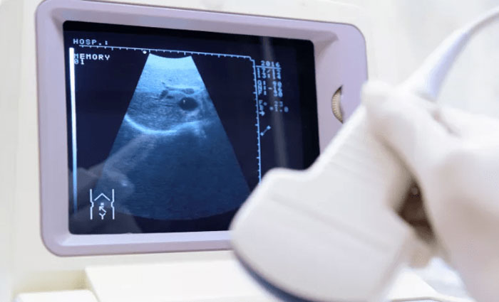
The issue lay in the limitations of B-ultrasound technology, typically only effective up to a depth of six or seven centimeters. Renea’s abdominal thickness was a staggering fifty to sixty centimeters, well beyond the ultrasound’s capacity. This meant that the doctors couldn’t accurately ascertain what was happening inside her abdomen using this method. They faced a diagnostic roadblock, complicating their efforts to understand and address Renea’s critical condition.